
Root canal transportation: from understanding to prevention
Most root canals are curved, whereas endodontic instruments are manufactured from straight metal blanks, resulting in root canal transportation. This may result in unprepared areas of the root canal wall and several distinct preparation errors (i.e., zip, elbow, ledging, perforation, strip perforation, outer widening, apical blockage, or damage to the apical foramen) that affects the treatment outcome.
The importance of root canal transportation is well illustrated by the number of publications evaluating the transportation of various instrumentation techniques with several experimental models using simulated canals or extracted teeth.
In clinical practice, root canal transportation can be minimized or avoided by understanding anatomy, adequate of X ray imaging, access cavity preparation and proper instrument selection and shaping technique.
In the lecture, the methods of transportation evaluation will be briefly outlined, followed by the clinical steps to minimize transportation, supported with clinical cases.
Ahmed HMA. 2022. A critical analysis of laboratory and clinical research methods to study root and canal anatomy. Int Endod J. 55(S2):229–280. doi:10.1111/iej.13702.
Fidler A, Plotino G, Kuralt M. 2021. A Critical Review of Methods for Quantitative Evaluation of Root Canal Transportation. J Endod. 47(5):721–731.
Hülsmann M. 2022. A critical appraisal of research methods and experimental models for studies on root canal preparation. Int Endod J. 55(S1):95–118. doi:10.1111/iej.13665.
Hartmann RC, Fensterseifer M, Peters OA, de Figueiredo JAP, Gomes MS, Rossi-Fedele G. 2019. Methods for measurement of root canal curvature: a systematic and critical review. Int Endod J. 52(2):169–180.
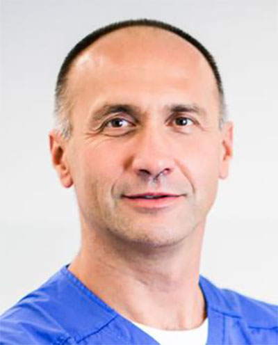
Currently he is a head of the Department of endodontic at University clinical Centre, Ljubljana Slovenia, full professor at Faculty of Medicine, University of Ljubljana, and head of the research group Periodontal Medicine. He was/is involved in the teaching of tooth anatomy, radiology, dental materials, and operative dentistry. In 2013, he was a visiting professor at Ege University, Izmir. He is a member of the Clinical practice committee at the European Society of Endodontology, He regularly lectures at national and international meetings. Since 2008 he works in part-time private dental practice, dedicated to endodontics.
His main areas of research are image analysis, dental materials and instruments. He applied his broad knowledge and experience to several areas of dental research, some of his findings proved to be universal and useful in medicine and general image analysis. He cooperates with researchers in the field of physics and technology (Elettra Sincrotrone Trieste Italy, Faculty of Electrical Engineering and Faculty of Mechanical Engineering of the University of Ljubljana). He has (co)authored 40 publications in indexed journal has over 340 citations (h index=11). He has co-authored 2 university textbooks.
Recently, he has been mainly involved in 3D image analysis obtained by μCT or the fusion of complementary modalities of clinical imaging techniques, namely CBCT and 3D optical scanning, which is used for the quantitative assessment of changes in hard and soft tissues in the oral cavity.
This site uses cookies. Find out more about cookies and how you can refuse them.
